
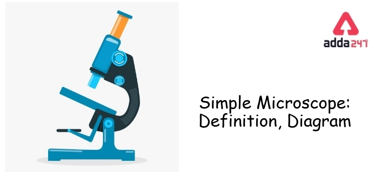
Simple Microscope
We have used various types of equipment in our laboratories. A simple Microscope is one of the most common and widely used equipment. There are many small objects which can’t be seen by the naked eye. The microscope is used for observing the magnified structure of those minute objects. Different types of microscopes are simple microscopes, compound microscopes, Light microscopes, Electron microscopes, etc. Simple Microscope plays a very significant role in various fields such as Biology labs, Medical Science, etc. The function of a simple Microscope is to provide a clear view of a small object or specimen in a magnified way. In this article, we are going to understand Microscope parts, diagrams, its work principle, and applications.
Simple Microscope Definition
A simple Microscope is an instrument with a specially modified magnifying glass that can magnify minute objects or specimens to make organisms visible to the eyes. There are different types of microscopes These are Simple Microscope has a bi-convex lens with a short focal length. Along with the lens, there are various parts in the microscope .lets understand the parts of a Microscope one by one.
Simple Microscope is Discovered By
The simple Microscope is discovered by Antonie van Leeuwenhoek and That’s why he is considered to be one of the first microbiologists.
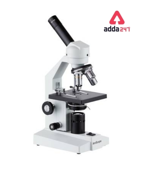
Simple Microscope Diagram
Simple Microscope Formula
Simple Microscope Formula is denoted as M = 1 + D/F, where D is the shortest distance of distinct vision and F is the focal length of the convex lens.
Noted Fact: The higher the magnifying power of the microscope, the shorter the focal length of the lens.
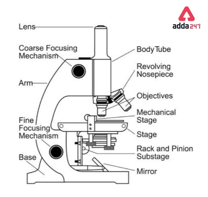
Simple Microscope Parts with Diagram
As we can see in the above image of the Microscope, there are various optical parts as well as mechanical parts of the Microscope. So we can classify the structure into two parts such as –
A. Mechanical Parts
The mechanical part is a framework to which various functional parts are attached.
• Base – Base provides Mechanical stability to the entire structure of the microscope.
• Piller – Two upright pillars project up from the base and are attached to the C-shaped handle. The hinge point allows the microscope to be at a suitable angle for comfortable viewing.
• Body tube- Fitted at the upper end of the handle either vertically or at the angle, the body tube is the part through which light passed to the eyepiece. It is 15 – 16 cm in length and can be raised or lowered by the focusing system.
•The Stage – Stage is two types – Fixed stage and Mechanical stage.
a) Fixed Stage– it is a square platform with an aperture in its center and fitted to the limb below the objective lens.
The side is placed on it and centered over the aperture for viewing.
b) Mechanical Stage –It is a calibrated metal frame fitted on the right edge of the fixed stage. there is a spring-mounted clip holding the slide in position.
• The focusing system: Course and fine adjustment knob
The focusing system consists of a course and a fine adjustment knob. There are two courses and two fine optical systems up or down rapidly through a large distance while the fine adjustment works the same way but moves
Course and fine optimal distance through a small distance.
B) The Optical parts
• The eyepiece – The eyepiece fits on top of the body tube . Most microscopes are provided with 5X,10X, and 15X eyepiece lenses.
• The Nose piece – It is fitted at the lower end of the body tube and has two parts:- The fixed nose piece and the revolving nose piece.
• Source of light- The light source may be outside the microscope (External light source ) or within an internal light source ( Internal light source)
• The mirror -In a simple Mirror is concave-reflecting type.
• The Condenser- The condenser is a system of lenses fitted in a short cylinder that is mounted below the stage. It can be raised or lowered by a rack and pinion and focuses the eight rays into a material under study
• The iris Diaphram- It is fitted within the condenser. A small lever on the side adjusts the size of the aperture of the diaphragm, thus allowing more or less light to follow on the material under study.
Simple Microscope Magnifying Power
Magnification is the ratio between the apparent size of the objects as seen in their virtual image and their actual size. It is given by the product of magnifications by the object and eyepiece lens.
For example,
With a 40X objective and a 10X eyepiece.The image gets magnified 40 X 10 =400 times .Magnification may be enhanced by increasing the power of either of both lens systems.
To determine the magnification power of a simple microscope,we can use the following equation:
M = 1 + D/F
[ Here D is represented as the shortest distance of the distinct vision and F is the focal length of the convex lens. ]
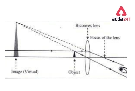
Simple Microscope: Principle with Ray Diagram
To obtain small magnifications, a simple microscope is used. It is typically used to investigate microscopic algae, fungi, and biological specimens. A semipermeable object passes light from a source of light (mirror) (Figure 1). A biconvex lens enlarges the virtual image by magnifying the element size. The image is seen from the opposite side.
Simple Microscope: Applications
Simple Microscope used widely in various fields –
A) In biology laboratories, a Simple Microscope is used to study minute organisms such as bacteria, viruses, algae, etc
B) Simple Microscope is also used in a pathology laboratory to examine blood, urine, etc.
C) Watchmakers also used simple Microscopes to see small watch parts in a magnified view.
Simple Microscope in Hindi
सरल माइक्रोस्कोप: हमने अपनी प्रयोगशालाओं में विभिन्न प्रकार के उपकरणों का उपयोग किया है। एक साधारण माइक्रोस्कोप सबसे आम और व्यापक रूप से उपयोग किए जाने वाले उपकरणों में से एक है। बहुत सी ऐसी छोटी-छोटी वस्तुएं हैं जिन्हें नंगी आंखों से नहीं देखा जा सकता है। सूक्ष्मदर्शी का उपयोग उन सूक्ष्म वस्तुओं की आवर्धित संरचना को देखने के लिए किया जाता है। विभिन्न प्रकार के सूक्ष्मदर्शी सरल सूक्ष्मदर्शी, यौगिक सूक्ष्मदर्शी, प्रकाश सूक्ष्मदर्शी, इलेक्ट्रॉन सूक्ष्मदर्शी आदि हैं। सरल सूक्ष्मदर्शी जीव विज्ञान प्रयोगशाला, चिकित्सा विज्ञान आदि जैसे विभिन्न क्षेत्रों में एक बहुत महत्वपूर्ण भूमिका निभाता है। एक साधारण सूक्ष्मदर्शी का कार्य एक स्पष्ट सूक्ष्मदर्शी प्रदान करना है। किसी छोटी वस्तु या नमूने को बड़े तरीके से देखना। इस लेख में, हम माइक्रोस्कोप भागों, आरेखों, इसके कार्य सिद्धांत और अनुप्रयोगों को समझने जा रहे हैं।
सरल माइक्रोस्कोप परिभाषा
एक साधारण माइक्रोस्कोप एक विशेष रूप से संशोधित आवर्धक कांच वाला एक उपकरण है जो आंखों को दिखाई देने वाले जीवों को बनाने के लिए सूक्ष्म वस्तुओं या नमूनों को बड़ा कर सकता है। विभिन्न प्रकार के सूक्ष्मदर्शी होते हैं ये साधारण सूक्ष्मदर्शी में लघु फोकल लंबाई के साथ एक द्वि-उत्तल लेंस होता है। लेंस के साथ-साथ सूक्ष्मदर्शी में विभिन्न भाग होते हैं। आइए सूक्ष्मदर्शी के भागों को एक-एक करके समझते हैं।
सरल सूक्ष्मदर्शी की खोज एंटोनी वैन लीउवेनहोएक ने की थी और यही कारण है कि उन्हें पहले सूक्ष्म जीवविज्ञानी में से एक माना जाता है।
Related Post:
- Fleming’s Left Thumb Rule Used For
- Cube Formula- Volume, Surface Area In Maths For Class 10
- Water Cycle Diagram For Kids Of Class 3 With Explanation
- 1 To 30 Square And Cube [ PDF Download ]
- Trigonometry Table (0 To 360): Formula, Value, Chart, Ratio, PDF For Class 10, 12
Simple Microscope Diagram: FAQs
Q. What is a Simple Microscope?
A simple Microscope is an instrument with a specially modified magnifying glass that can magnify minute objects or specimens to make organisms visible to the eyes. These are Simple Microscope has a bi-convex lens with a short focal length.
Q. What is the working principle of a simple Microscope?
To obtain small magnifications, a simple microscope is used. It is typically used to investigate microscopic algae, fungi, and biological specimens. A semipermeable object passes light from a source of light (mirror). A biconvex lens enlarges the virtual image by magnifying the element size. The image is seen from the opposite side.
Q. How to calculate the magnification power of a simple Microscope?
The magnification power of a simple microscope can be calculated by the following equation. M = 1 + D/F
[ Here D represented the shortest distance of the distinct vision and F is the focal length of the convex lens. ]
Q. Write down the two applications of Simple Microscope?
Two applications of the Simple Microscope are- A) In biology laboratories, the Simple Microscope is used to study minute organisms such as bacteria, viruses, algae, etc, B) the Simple Microscope is also used in a pathology laboratory to examine blood, urine, etc.


 CLAT Logical Reasoning Preparation Tips
 How to Prepare Current Affairs for CLAT
 Best Strategy to Score High in CLAT Lega...