
Have you ever considered the origin of urine? That fluid from your body that consistently forces you to go to the restroom? The kidney generates urine, which consists of water, urea, and salts. While blood flows through the kidney, filtration and selective reabsorption take place, resulting in urine as the final product. In the kidney, numerous small tubules are involved in the process of urine creation. These structures are known as nephrons.
Let’s examine the structure and function of the nephron and explore in depth the various components of the nephron and the role of the renal tubule in the processes of urine formation and excretion.
Nephron
The structure that really makes urine in the process of eliminating waste and excess substances from the circulation is called a Nephron. Each human kidney contains approximately 1,000,000 Nephrons. The most rudimentary Nephrons can be found in primitive fish kidneys (pronephros), amphibian larvae, and embryos of more evolved vertebrates.
The Nephrons seen in amphibians’ and most fish’s kidneys (mesonephros), as well as in the late embryonic development of more sophisticated vertebrates, are only marginally more advanced in structure. Adult kidneys, or metanephros, of land vertebrates such as reptiles, birds, and mammals have the most mature nephrons.
Definition of Nephron
A nephron serves as the fundamental structural and functional component of the kidney. They are the tiny structures made up of a renal corpuscle and a renal tubule. The term nephron originates from the Greek term – nephros, which signifies kidney. Each human kidney contains around millions of nephrons. These structures are one of the most fundamental concepts that every student needs to study in Biology.

Nephron Diagram
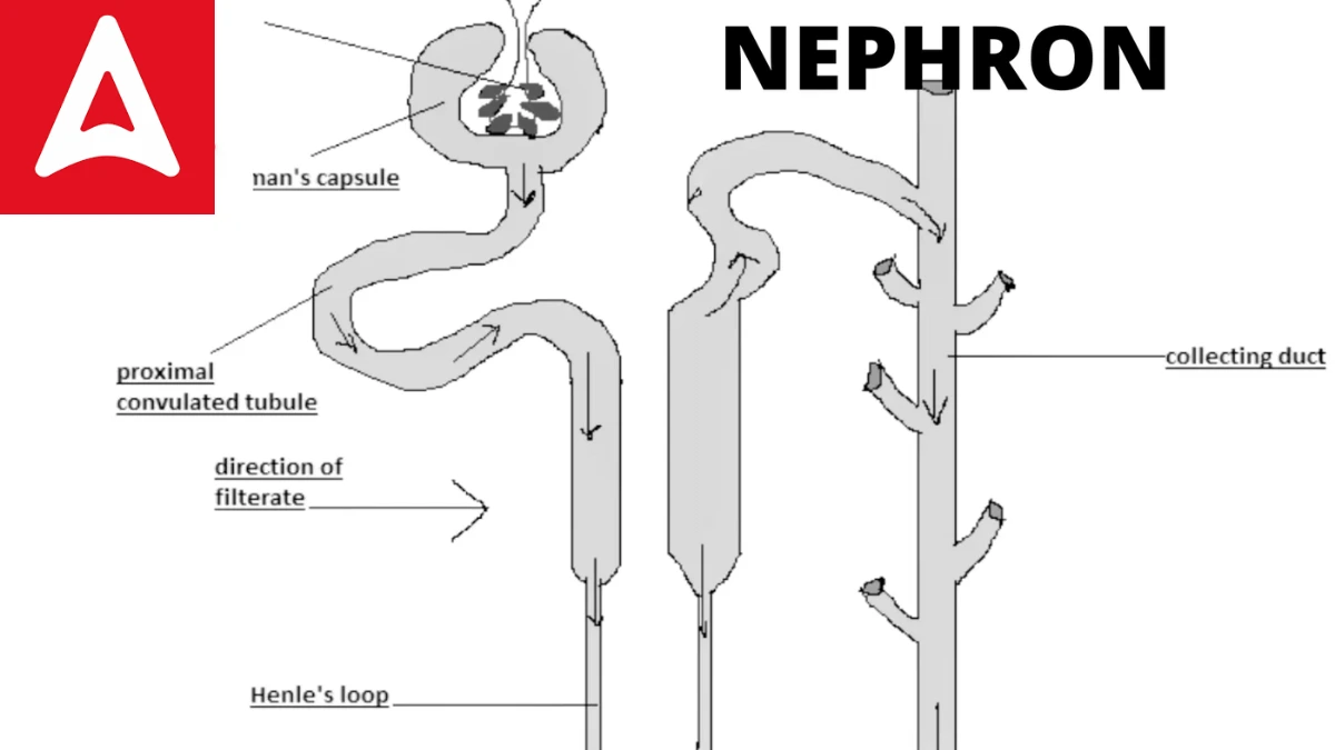
The diagrammatic representation of Nephron is given below:
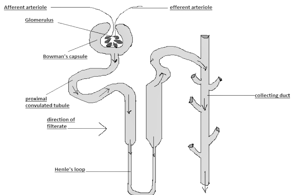
Figure: Diagrammatic Representation of Nephron
Nephron Structure
In the mammalian kidney, each nephron is a long tubule, or exceedingly fine tube, measuring 30–55 mm (1.2–2.2 inches) in length. This tube is closed, inflated, and folded into a cuplike shape with two walls. The structure of nephron comprises two major portions: Renal Tubule and Renal Corpuscle
The renal corpuscular capsule, also known as Bowman’s capsule, encloses the glomerulus, a cluster of small blood arteries called capillaries. The renal corpuscle is made up of the capsule and glomerulus. Blood travels into and out of the glomerulus via tiny arteries known as arterioles, which enter and exit the glomerulus through the capsule’s open end.
Fluid filters out of the blood in the glomerulus via the inner wall of the capsule and into the nephron tubule in the renal corpuscle. The secretion of certain compounds into the filtrate and the selective reabsorption of water and other ingredients from it change its composition as it moves through the tubule. Urine is the final product, which is transported into the renal pelvis via the collecting tubules.
Structure of Nephron in Detail
The nephron is the functional unit of the kidney, responsible for filtering the blood and producing urine. structure of nephron consists of several distinct regions and structures that work together to perform the filtration, reabsorption, and secretion processes. The structure of nephron can be divided into the following main parts:
- Renal Corpuscle: The renal corpuscle consists of two main components:
- Glomerulus: It is a network of tiny capillaries where blood filtration occurs. The glomerulus is surrounded by a cup-like structure called Bowman’s capsule.
- Bowman’s Capsule: It encloses the glomerulus and collects the filtrate from the blood.
- Proximal Convoluted Tubule (PCT): The filtrate from Bowman’s capsule enters the PCT, which is a twisted tubular structure. The PCT is responsible for the reabsorption of water, ions, glucose, amino acids, and other important substances from the filtrate back into the bloodstream.
- Loop of Henle: The Loop of Henle is a U-shaped tube that descends into the medulla of the kidney and then ascends back towards the cortex. It consists of a descending limb and an ascending limb. The Loop of Henle plays a crucial role in creating concentration gradients within the kidney, allowing for the reabsorption of water and electrolytes.
- Distal Convoluted Tubule (DCT): The DCT is another twisted tubular structure that follows the Loop of Henle. It is responsible for fine-tuning the reabsorption and secretion processes. It helps regulate the pH and electrolyte balance of the filtrate.
- Collecting Duct: Multiple DCTs merge into a larger collecting duct, which passes through the medulla of the kidney. The collecting duct receives filtrate from several nephrons and carries it towards the renal pelvis. The collecting duct plays a role in the final concentration and volume adjustments of urine.
- Renal Pelvis: The renal pelvis is a funnel-shaped structure that collects urine from the collecting ducts. It serves as a pathway for urine to flow from the kidney to the ureter and eventually out of the body.
It’s important to note that there are millions of nephrons present in each kidney, and they work collectively to filter and process blood continuously, maintaining the body’s fluid and electrolyte balance.
The structure of nephron allows for the filtration of blood, reabsorption of essential substances, and the excretion of waste products to produce urine. This intricate system ensures the proper functioning of the kidneys and the maintenance of homeostasis in the body.
Function of Nephron
The function of Nephron is dependent on its two major parts:
Renal Tubule
The renal tubule is a lengthy, convoluted structure that arises from the glomerulus and is separated into three functional segments. Because of its proximity to the glomerulus, the initial section is called the proximal convoluted tubule (PCT), and it remains in the renal cortex. Because it creates a loop (with descending and ascending limbs) that travels through the renal medulla, the second part is called the loop of Henle, or nephritic loop. The distal convoluted tubule (DCT) is the third portion of the renal tubule, and it is similarly limited to the renal cortex.
Bowman’s capsule is a cup-like structure that surrounds the glomerulus’ capillaries. This structure continues to generate PCT tubules, which are highly coiled tubules. PCT continues to form the Henle loop, which leads to DCT, which then leads to the collecting duct. Tubules’ primary role is reabsorption, which can be accomplished through active or passive transport. Tubule secretions also aid in the production of urine without disrupting the body’s electrolyte balance.
Renal Corpuscle
A glomerulus is enclosed by a Bowman’s capsule in the renal corpuscle. An afferent arteriole gives rise to the glomerulus, which then empties into an efferent arteriole. The smaller diameter of an efferent arteriole aids in the maintenance of high glomerular blood pressure. There are three levels to Bowman’s capsule:
- The outer parietal layer is made up of epithelial cells with tiny holes of 12nm in width.
- This stratum of the basement membrane is selectively permeable.
- The inner visceral layer is made up of big nucleated cells called podocytes that have podocel, which are finger-like projections.

Nephron Kya Hai in Hindi
नेफ्रॉन क्या है?
संचलन से अपशिष्ट और अतिरिक्त पदार्थों को हटाने की प्रक्रिया में वास्तव में मूत्र बनाने वाली संरचना को नेफ्रॉन कहा जाता है। प्रत्येक मानव गुर्दे में लगभग 1,000,000 नेफ्रॉन होते हैं। सबसे अल्पविकसित नेफ्रॉन आदिम मछली के गुर्दे (प्रोनफ्रोस), उभयचर लार्वा और अधिक विकसित कशेरुकियों के भ्रूण में पाए जा सकते हैं। उभयचरों और अधिकांश मछलियों के गुर्दे (मेसोनेफ्रोस) में देखे जाने वाले नेफ्रॉन, साथ ही अधिक परिष्कृत कशेरुकियों के देर से भ्रूण के विकास में, संरचना में केवल मामूली रूप से अधिक उन्नत होते हैं। सरीसृप, पक्षियों और स्तनधारियों जैसे भूमि कशेरुकियों के वयस्क गुर्दे, या मेटानेफ्रोस में सबसे परिपक्व नेफ्रॉन होते हैं।
नेफ्रॉन संरचना
स्तनधारी गुर्दे में, प्रत्येक नेफ्रॉन एक लंबी नलिका या अत्यधिक महीन ट्यूब होती है, जिसकी लंबाई 30-55 मिमी (1.2-2.2 इंच) होती है। यह ट्यूब बंद है, फुलाया जाता है, और दो दीवारों के साथ एक कप के आकार में मुड़ा हुआ है। नेफ्रॉन की संरचना में दो प्रमुख भाग होते हैं: वृक्क नलिका और वृक्क कोषिका
रीनल कॉर्पसकुलर कैप्सूल, जिसे बोमन कैप्सूल के रूप में भी जाना जाता है, ग्लोमेरुलस को घेरता है, छोटी रक्त धमनियों का एक समूह जिसे केशिकाएं कहा जाता है। वृक्क कोषिका कैप्सूल और ग्लोमेरुलस से बनी होती है। रक्त ग्लोमेरुलस में और बाहर छोटी धमनियों के माध्यम से यात्रा करता है जिन्हें धमनी के रूप में जाना जाता है, जो कैप्सूल के खुले सिरे के माध्यम से ग्लोमेरुलस में प्रवेश और बाहर निकलते हैं। द्रव ग्लोमेरुलस में रक्त से बाहर कैप्सूल की भीतरी दीवार के माध्यम से और वृक्क कोषिका में नेफ्रॉन नलिका में फ़िल्टर करता है। छानने में कुछ यौगिकों का स्राव और पानी और अन्य अवयवों के चयनात्मक पुनर्अवशोषण इसकी संरचना को बदल देते हैं क्योंकि यह नलिका के माध्यम से चलता है। मूत्र अंतिम उत्पाद है, जिसे एकत्रित नलिकाओं के माध्यम से वृक्क श्रोणि में ले जाया जाता है
नेफ्रॉन समारोह
नेफ्रॉन का कार्य इसके दो प्रमुख भागों पर निर्भर करता है:
वृक्क नलिका
वृक्क नलिका एक लंबी, जटिल संरचना है जो ग्लोमेरुलस से निकलती है और तीन कार्यात्मक खंडों में विभाजित होती है। ग्लोमेरुलस से इसकी निकटता के कारण, प्रारंभिक खंड को समीपस्थ घुमावदार नलिका (पीसीटी) कहा जाता है, और यह वृक्क प्रांतस्था में रहता है। क्योंकि यह एक लूप (अवरोही और आरोही अंगों के साथ) बनाता है जो वृक्क मज्जा के माध्यम से यात्रा करता है, दूसरे भाग को हेनले का लूप, या नेफ्रिटिक लूप कहा जाता है। डिस्टल कन्फ्यूज्ड ट्यूब्यूल (DCT) वृक्क नलिका का तीसरा भाग है, और यह इसी तरह वृक्क प्रांतस्था तक सीमित है।
बोमन कैप्सूल एक कप जैसी संरचना है जो ग्लोमेरुलस की केशिकाओं को घेर लेती है। यह संरचना पीसीटी नलिकाओं को उत्पन्न करना जारी रखती है, जो अत्यधिक कुंडलित नलिकाएं हैं। पीसीटी हेनले लूप बनाना जारी रखता है, जो डीसीटी की ओर जाता है, जो बाद में एकत्रित वाहिनी की ओर जाता है। नलिकाओं की प्राथमिक भूमिका पुनर्अवशोषण है, जिसे सक्रिय या निष्क्रिय परिवहन के माध्यम से पूरा किया जा सकता है। ट्यूबल स्राव शरीर के इलेक्ट्रोलाइट संतुलन को बाधित किए बिना मूत्र के उत्पादन में भी सहायता करते हैं।
गुर्दे की कणिका
एक ग्लोमेरुलस वृक्क कोषिका में एक बोमन कैप्सूल से घिरा होता है। एक अभिवाही धमनी ग्लोमेरुलस को जन्म देती है, जो फिर एक अपवाही धमनी में खाली हो जाती है। एक अपवाही धमनी का छोटा व्यास उच्च ग्लोमेरुलर रक्तचाप के रखरखाव में सहायता करता है। बोमन कैप्सूल के तीन स्तर हैं:
बाहरी पार्श्विका परत 12nm चौड़ाई के छोटे छिद्रों वाली उपकला कोशिकाओं से बनी होती है।
तहखाने की झिल्ली का यह स्तर चुनिंदा पारगम्य है।
आंतरिक आंत की परत पोडोसाइट्स नामक बड़ी न्यूक्लियेटेड कोशिकाओं से बनी होती है, जिसमें पोडोकेल होता है, जो उंगली की तरह के प्रोजेक्शन होते हैं।
Related Post:
- Important Question Of Physics Class 12 With Answers
- Differentiation: Equations, Formula, Sum, Product, Quotient And Chain Rule
- Job Application- Sample Letter, Format, Examples, Biodata
- Inches To Centimeters- Conversion, Chart & Formula
- Noun: Definition, Types, And Examples
- Formal Letter Format And Examples
- Acute Angle: Definition, Degree, Triangle, Example
- Reactivity Series Of Metals And Non-Metals


 Breathing and Exchange of Gases Notes fo...
 Omnivores Animals- Definition, Name List...
 Life Processes: Check Nutrition, Transpo...