
Layers of Retina
To know about Layers of Retina first, we know about Retina. For most vertebrates and some molluscs, the retina is the innermost, light-sensitive layer of tissue in the eye. The retina receives a focused two-dimensional image of the visual world from the eye’s optics, analyses it there, and sends nerve impulses carrying that processed image to the visual cortex via the optic nerve to produce vision. The retina performs a function that is similar to the film or image sensor of a camera in many respects. Layers of Retina details are given below.
Layers of Retina Histology
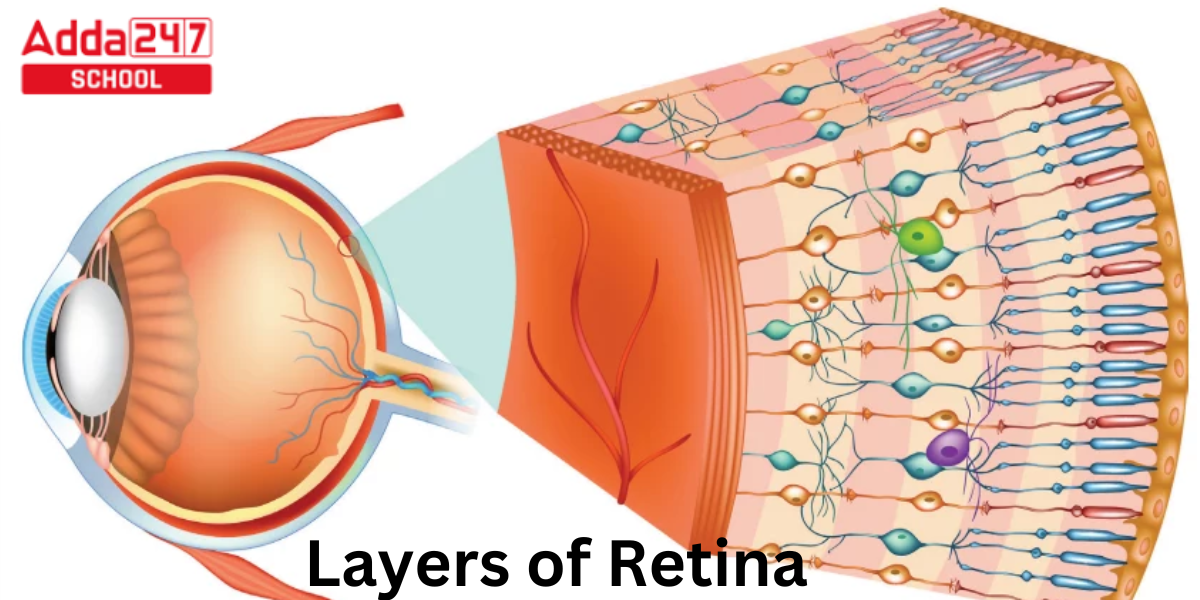
Layers of the Retina have been discussed here. The retina of a vertebrate comprises ten different layers. From the vitreous body outward, in order of proximity:
- Müller cells create the inner limiting membrane and basement membrane.
- A thin layer of Müller cell footplates is present between the nerve fibre layer and the inner limiting membrane, which can be seen as the axons of the ganglion cell bodies.
- The ganglion cell layer is composed of the nuclei of ganglion cells, whose axons develop into the fibres of the optic nerve, as well as a few dislocated amacrine cells.
- The synapse between the axons of the bipolar cell and the dendrites of the ganglion and amacrine cells is located in the inner plexiform layer.
- Amacrine cells, bipolar cells, and horizontal cells’ nuclei and surrounding cell bodies are found in the inner nuclear layer.
- Outer plexiform layer – rod and cone projections that join the dendrites of horizontal and bipolar cells at the rod spherule and cone pedicle, respectively. This is referred to as the Fiber layer of Henle in the macular area.
- Rods and cones’ cell bodies make up the outer nuclear layer.
- The layer that divides the photoreceptors’ inner segment components from their cell nuclei is known as the external limiting membrane.
- Rods and cones are divided between inner and outer segments, with the outer segments containing a highly sophisticated light-sensing device.
- One layer of cuboidal epithelial cells makes up the retinal pigment epithelium. The neural retina is nourished and supported by this layer, which is closest to the choroid. The black pigment melanin in the pigment layer reduces light reflection around the globe of the eyeball, which is crucial for good vision.
Know All About Human Eye
Layers of Retina Diagram
Layers of Retina Physiology
- Photoreceptor cell
- Bipolar cell
- Retinal ganglion cell
Layers of Retina Anatomy Anterior to Posterior
Layers of Retina- The human retina has a surface area of around 1,100 square millimetres and measures 32 millimetres from ora to ora along the horizontal meridian. 2 The retina has an average thickness of 200 micrometres; it is slightly thicker between the macula and the optic nerve head and gradually becomes thinner at the ora serrata and the fovea.
Area centralis
The retina’s area centralis is a circular region with a diameter of 5–6 mm. It is located between the superior and inferior temporal arteries.
Equator
The fundus’s equator is located 14 to 15 mm or so away from the limbus. Finding the vortex veins is clinically one of the simplest ways to detect the fundus’ equator. In each of the eye’s four quadrants, the choroidal veins empty into a vortex vein. There are typically four vortex veins (one in each quadrant), but there could be up to ten.
Peripheral Retina
The remaining retina is referred to as the peripheral retina. On the temporal side, the distance from the optic disc to the ora serrata is 23–24 mm, while on the nasal aspect, it is roughly 18.5 mm. The retina’s thickness ranges from 110 to 140 m in the peripheral region, and increases as it moves towards the posterior pole.
Cellular Structures in Retina
The three types of neurons that make up the majority of the neurosensory retina are photoreceptors, bipolar cells, and ganglion cells. Amacrine cells and horizontal cells, two more significant neurons, play supporting roles. In a three-step journey through the retina, the photoreceptor cells, bipolar cells, and ganglion cells carry the neurological signal. Sensory receptors include photoreceptors. Ganglion cells create second-order neurons, while bipolar cells are first-order cells.
Retinal Pigment Epithelial Cells (RPE cells)
The RPE cell is a cuboidal cell with a hexagonal shape that is located behind the photoreceptors. Between Bruch’s membrane and the neurosensory retina, a single layer of RPE cells that are joined to one another along their sides. The photoreceptor tip is encircled by microvilli on the apical surface of the RPE cell.
Photoreceptors
The unique sense cells known as photoreceptors can absorb light photons and contain photopigments. The rods and the cones are the two different types of photoreceptors. Rods sense contrast, brightness, motion, and typically form black-and-white images, which are necessary for vision in low light or complete darkness. Cones are in charge of colour vision, fine resolution, spatial resolution, and vision under strong light. Varied regions of the retina have different densities of rods and cones. The number of rods increases toward the periphery before again dropping toward the extreme periphery, where they are nonexistent at the fovea. Nearer the macula, the cone density rises, and there are only cones in the foveal region.
|
Rod |
Cone |
| There are between 110 and 125 million rods in the human retina overall, according to estimates. | Cones in the human retina are thought to total between 6.3 and 6.8 million. |
| Rods are in charge of detecting brightness, contrast, and motion. | Cones are in charge of colour vision, spatial resolution, and fine resolution. |
| Rods are absent at fovea | Cones are most dense at fovea |
| The number of rods increases towards the periphery | Number of cones decreases towards periphery |
| The outer segment is rod-shaped | The outer segment is cone-shaped |
| Only one type, monochromatic vision | Three types, color vision |
| Contains Rhodopsin | Contain Photopsin |
| Regenerative power is high | Regenerative power is relatively low |
| Smaller in Size | Relatively Larger |
| Slower response speed | Faster response |


 CLAT 2027 Preparation: Complete Study Pl...
 Best CLAT Online Course 2027: Why Adda24...
 Best Strategy to Attempt the CUET Exam f...