
Fluid Mosaic Model: S.J. Singer and Garth L. Nicolson initially proposed the fluid mosaic model in 1972. This widely regarded Fluid Mosaic Model explains the structure of animal cell plasma membranes. According to this hypothesis, membrane components such as proteins and glycolipids form a mobile mosaic in the fluid-like environment formed by an ocean of phospholipids. Isn’t the Fluid Mosaic Model’s structure interesting? It might also be a fun science project for your school’s model competition. In this article, we will learn about the structure of this membrane model and how to create a realistic fluid mosaic model at home.
Fluid Mosaic Model
The lipid bilayer model was proposed more than 25 years ago. It all started with the idea that the membrane was formed of a lipid bilayer, in which membrane phospholipid developed into a dual layer with non-polar, hydrophobic tails towards each other. The hydrophilic ‘head’ sections are exposed to the cytosol and extracellular area.
In the 1950s, cell membranes were first visualized. Initially, it appeared that the lipid membrane had been sealed on both sides by thin sheets of protein. However, two scientists, Singer and Nicolson, developed this concept in 1972 to establish the fluid mosaic model.
The fluid feature of the plasma membrane is due to its construction as a mosaic of components – phospholipids, cholesterol, proteins, and carbohydrates. The ratios of proteins, lipids, and carbohydrates in the cell’s plasma membrane differ depending on the cell type. Myelin, for example, is composed of 18% protein and 76% lipid. The inner mitochondrial membrane is 76% protein and 24% lipid.
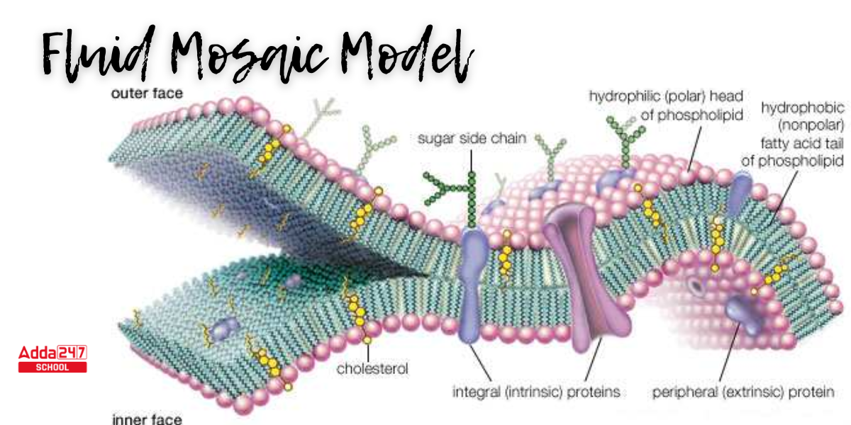
Fluid Mosaic Model Diagram
The fluid mosaic model has altered slightly over time, but it currently perfectly reflects the plasma membrane’s structure and functions as we presently understand them. The fluid mosaic model diagram represents the lipid bilayer with various types of integral membrane proteins, in addition to cholesterol, glycoproteins, and glycolipids. The image also depicts the membrane’s attachment to the cytoskeleton.
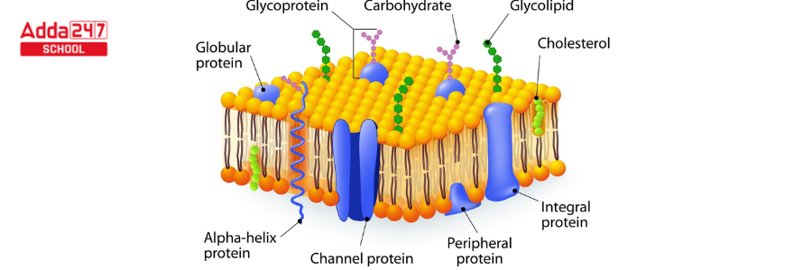
Fluid Mosaic Model of Plasma Membrane
The fluid mosaic model explains Human red blood cells, which can be seen under a light microscope, are around 8 m broad, or 1,000 times bigger than a plasma membrane. The Fluid Mosaic Model of plasma membrane structure is discussed below.
- The membrane is fluid, but it also has an inherent structure, as it is attached to the cytoskeleton, according to Singer and Nicolson’s Fluid Mosaic Model of plasma membrane.
- An experiment in which membranes of different components were artificially united first demonstrated the fluid nature of the lipid matrix creating the membrane. In less than an hour, the proteins from both cells redistributed themselves throughout the entire joined membrane.
- In the Fluid Mosaic Model, the phospholipid bilayer was supposed to be punctuated by numerous proteins that generated a mosaic-like pattern in the lipid membrane.
- These proteins have the ability to cross the whole membrane or connect to one of the two lipid layers. Some proteins may even be linked to the membrane by a short lipid chain alone.
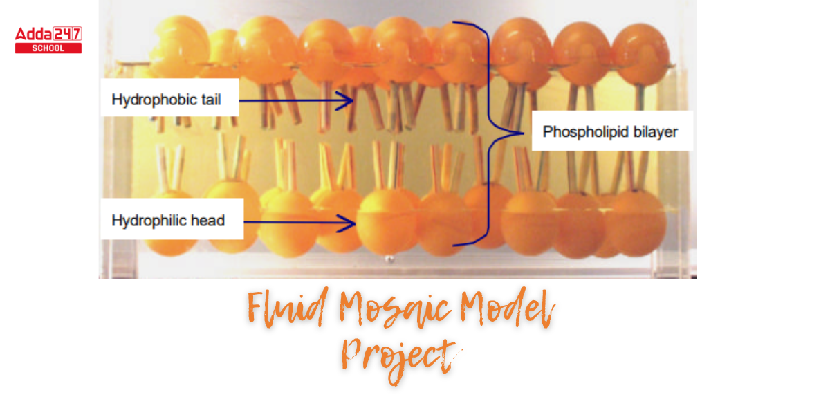
Fluid Mosaic Model of Cell Membrane Project
The fluid mosaic concept can be displayed with a fluid Mosaic model composed of low-cost components that are easily available. All of the materials used in this fluid mosaic model project are affordable and can be obtained from any stationary store.
Material Required for a Fluid Mosaic Model
Students must have the following items before beginning the fluid mosaic model project.
- Ping-pong balls,
- drinking straws,
- a plastic container,
- oil
- water
- glue gun or normal glue
Instruction
Students need to follow the step-by-step process accurately to successfully complete the Fluid Mosaic Model.
Step 1. At first, cut the straws into 2-3cm long pieces.
Step 2. Using a glue gun, attach two drinking straw segments to each of the ping-pong balls.
Step 3. Separate the ping-pong balls into two groups. The phospholipids of the top layer are represented by one group, while the phospholipids of the lower layer are represented by the other.
(Note – The quantity of ping-pong balls used is determined by the size of the container.)
Step 4. Attach some weights (plasticine) to the open end of the straws you use for the upper set of ping-pong balls.
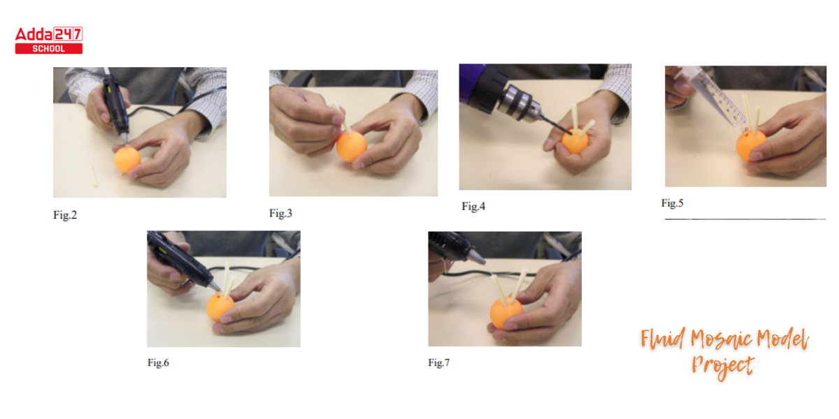
Step 5. Pierce a tiny hole in the ping-pong ball for the lowest set of ping-pong balls (Fig.4). Refill the ping-pong ball with water (Fig.5) and use the glue gun to seal it (Fig.6).
Step 6. Then, using the glue gun, close the open end of the drinking straw (Fig.7), so that every drinking straw becomes entangled with air. Fig.4
Step 7. Add the ingredients in the following order: salt water, lower group of ping pong balls, oil, and upper group of ping pong balls.
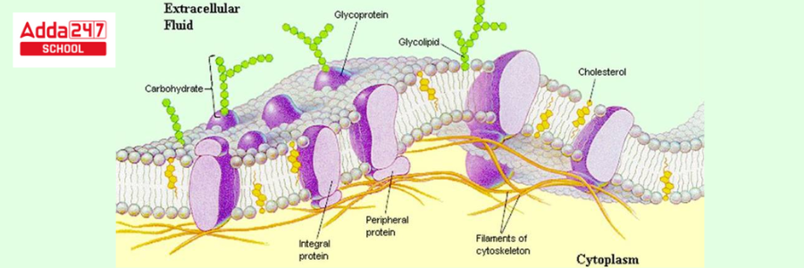
Explain Fluid Mosaic model of Plasma Membrane
We have explained all the components we have shown in the Fluid Mosaic Model of plasma membrane project here.
Phospholipids
A phospholipid molecule is made up of a three-carbon glycerol backbone, two molecules of fatty acids connected to carbons 1 and 2, and a group that contains phosphate attached to carbon 3. Phospholipids are amphipathic molecules that have a polar, hydrophilic ‘head’ region created by a phosphate group and a non-polar, hydrophobic ‘tail’ area composed of two long-chain fatty acids. A covalent connection holds them to a glycerol molecule.
Cholesterol
Although cholesterol is present in the phospholipid layer, the membrane retains its permeability and durability. It is found between the phospholipids and inhibits hydrophilic tails from compacting at low temperatures and expanding at high temperatures. Small molecules, such as carbon dioxide and oxygen, are able to pass easily across the membrane, whereas the cell maintains selective permeability for bigger molecules.
Proteins
There are three types of proteins in the plasma membrane:
Integral protein – Integral protein occupies the entire membrane, typically with alpha-helices constituting the transmembrane area. These proteins produce channels that allow big molecules and ions to flow over the membrane’s hydrophobic layer. Proteins could be embedded in a single leaflet of the membrane. These proteins are frequently used in signaling cascades and can operate as carrier molecules, generating a signal from one section of the membrane to another.
Peripheral proteins – Peripheral proteins are found concealed within a single membrane leaflet. They transport signals from one part of the membrane to another. Only a small lipid tail is inserted into the hydrophobic region of this protein, which is very loosely connected to the membrane.
Glycoproteins: Some membrane proteins can be covalently linked to carbohydrates to produce glycoproteins. These have the ability to interact with water molecules. They are in charge of membrane stabilization and intercellular communication. These have the ability to interact with water molecules. They develop hormone and neurotransmitter receptors.


 Best CLAT Coaching Classes in Rajouri Ga...
 IPMAT Coaching in Rajouri Garden Delhi |...
 CUET Coaching in Rajouri Garden Delhi | ...