
Compound Microscope: We’ve all seen compound microscopes in the school lab. A compound microscope is an exceptionally powerful microscope that uses a compound lens arrangement to see objects that our naked eyes cannot see. It is often used in laboratories to analyze specimens. A compound microscope includes multiple lenses, One is the objective lens which is typically 4x, 10x, 40x, or 100x is multiplied by the eyepiece lens (often 10x) to achieve magnifications of 40x, 100x, 400x, or 1000x. The resolution of a compound microscope is better, and the two sets of lenses produce a two-dimensional view of the sample. In this article, we are going to discuss how every compound microscope parts, how it works, the magnification, uses of compound microscope, and more.
Compound Microscope
The expression compound implies the use of multiple lenses in a microscope. The difference between a simple and complex microscope is that a simple microscope only has one lens, but a compound microscope has multiple lenses. Instead of a single magnifying lens, two lenses are used to achieve greater magnification. While the eyepieces and objective lenses provide great magnification, a condenser below the stage directs light into the sample.
The objective has a short focus length and is placed near the thing under examination. It is utilized to create a true image on the front focal plane of the second lens, often known as the eyepiece or ocular. The eyepiece creates an expanded virtual image that the spectator can view. The magnification of the compound microscope is the sum of the magnifications of the objective lens and the eyepiece.
Compound Microscope Discovered by
The design and development of the compound microscope by the Dutch optician Hans Jansen and his son Zacharias towards the end of the 16th century were some of the most significant improvements in diagnosis. In the late 16th century, a Dutch father-son pair named Hans and Zacharias Janssen constructed the first compound microscope when they realized that putting a lens at the top and bottom of a tube and looking through it magnified objects on the other end. Galileo, an Italian philosopher, astronomer, and mathematician, built a microscope and a telescope in the early 17th century. The utility of microscopes in biological sciences and diagnostics was first recognized in the late 17th century when Dutch microscopist Antonie van Leeuwenhoek was the very first to view protozoa and bacteria and one first to lay out red blood cells -erythrocytes.
Compound Microscope Diagram
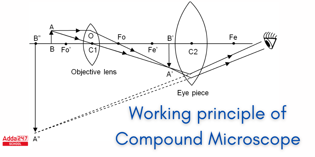
Multiple converging lenses can be used to achieve higher magnification. When two convex lenses are combined, the magnification is multiplied. This is referred to as a compound microscope. The operating compound microscope labeled diagram is shown below.

Working principle of Compound Microscope
As indicated in the diagram, the lens closest to the item is referred to as the objective, and it produces a true, inverted, enlarged representation of the object. This image is the subject of the second lens. The lens used to examine the final image is known as an eyepiece, and it acts similarly to a simple microscope or magnification. It generates an expanded and simulated image. The first inverted picture will be inside the focal distance when the final image is created at a close location. The initial inverted picture will be at focus distance when the final result is formed at infinity. In comparison to the original object, the resulting image is inverted.
Types of Compound Microscopes
Compound Microscopes can be divided into two distinct categories –
- Light Microscope
- Electron Microscope
Light Microscope
There are four types of light compound microscopes.
- Bright-Field Microscope: It employs an intense field illumination approach to produce a dark photograph of an object over a bright background. It is used to examine stained biological materials, tissues, and cells.
- Dark-Field Microscope: This microscope generates an illuminated picture of an object against a backdrop of darkness. It is used to observe live, unstained materials that include bacteria or tiny creatures.
- A phase-contrast microscope: It is used to examine clear objects that do not absorb or deflect light, such as biological cells or microbes.
- Fluorescent Microscope: This type of microscope uses a particular light source to stimulate molecules that are fluorescent in the sample. It is used to examine fluorescently labeled samples such as tissues, cells, or proteins.
Electron Microscope
There are three types of electron compound microscopes.
- Scanning Electron Microscope (SEM): Using a focused stream of electrons, this type of microscope creates incredibly detailed images of a specimen’s surface. The laser scans the specimen’s surface, producing an intricate 3D image.
- Transmission Electron Microscope (TEM): This particular kind of microscope creates a picture by passing an electron beam through a thin portion of the specimen. It generates precise photographs of the interior structure of cells, tissues, and various other biological samples at high resolution.
- Confocal Microscope: It scans the specimen at various depths with a laser beam, and a computer builds a three-dimensional model of the specimen from the acquired data. This sort of microscope is utilized to create three-dimensional images of the specimen.
Compound Microscope Parts
A compound microscope’s parts are divided into two types.
- Non-optical features
- Parts of optics
Non-optical Features
- Base – The base, often called the foot, can be alternatively U-like or horseshoe-shaped. It is a metal arrangement that holds the entire microscope together.
- Pillar – The pillar allows for a link between the bottom and the arm.
- Arm – The arm, also known as the limb, is a metallic handle that connects the arm to the inclined joint. The arm holds up the stage and the body tube.
- Joint Inclination – If the observation must be performed while sitting, the magnifying glass can be tilted through the inclination joint.
- Stage -It is the iron platform with an aperture in the center that is attached to the bottom half of the arm. To secure tiny slides to the stage for inspection, side clips or manual stage clips are utilized.
- Body Tube – The body tube’s principal function is to retain the objective and ocular lenses at both ends. The head is the end with the ocular lens, while the nose piece is the end containing the objective lens.
- Drawtube – The drawtube is a small fixed tube located at the top of the body tube. The major function of the drawtube is to support the ocular lens.
- Rack and Pinion -The rack and pinion are connected to the body tube or the stage so they bring the item into focus.
- Automatic Stop – A tiny screw on the rack and pinion prevents the body tube from slipping downhill. This protects the objective lens from harm.
- Adjustment Screws – There are two sets of adjusting screws, one for coarse adjustment and one for fine adjustment. Fine adjustments raise the body tube or stage very quickly, while coarse adjustments raise the body tube and stage. Careful editing can result in a clean image.
Compound Microscope Parts and Functions
Now we will discuss about the optical parts of a compound microscope.
Optical Components
- Diaphragm- The diaphragm controls the quantity of light that falls on an object. It can be found beneath the stage. The two distinct kinds of diaphragms are the disc and the iris.
Condenser: This is located beneath the diaphragm. Light can be focussed by adjusting the condenser, which can be adjusted up and down. - Reflector – A reflector is a mirror that is mounted above the base. The mirror features a plane mirror on one side and a concave mirror on the other. The plane mirror side is utilized when the light is strong, while the concave mirror side is employed when the light is weak.
- Objective lenses – These are located above the nosepiece. The objective lens is a compound lens that produces a true inverted image of the image contained within the body tube. Objective lenses are classified into two or three types: low power, high power, and oil immersion.
- Ocular Lens/Eyepiece – Another name for the ocular lens is the eyepiece. These lenses can magnify the image of minuscule objects. There are four types of magnification that can occur in the ocular lens: 5X, 10X, 15X, and 20X.
Magnification of Compound Microscope
A compound microscope is an erect microscope that combines two sets of lenses to achieve greater magnification than a simple or stereo microscope.
- Small test specimens that cannot be seen with the naked eye are viewed through compound microscopes. These samples are normally placed on a microscope slide. There is more room beneath the microscope for larger items such as rocks or flowers when employing a stereo microscope, and slides are not necessary.
- A two-dimensional image is produced by a compound microscope, but a three-dimensional image is produced by a stereo microscope.
- Compound microscopes normally have magnifications ranging from 40x to 1000x, whilst stereo microscopes have magnifications ranging from 10x to 40x.
- The compound microscope works on the basis that the combination of lenses increases the clarity of the sample. The material is initially observed in the tube as a primary picture, then in the eyepiece.
Advantages and Disadvantages of Compound Microscope
Since its development, the microscope has assisted many students and scientists in discovering and expanding our knowledge in basic biology, biomedical research, and medical diagnostics. Many advancements in microscopy techniques and lens engineering have contributed to the development of today’s microscope. Let us look at some of the advantages and disadvantages of compound microscopes.
Advantages of Compound Microscope
1) Simple to use.
2) Low cost in comparison to electron microscopes.
3) Capable of seeing live samples.
4) Capable of magnifying up to 2000 times.
Disadvantages of Compound Microscope
1) It is not possible to magnify more than 2000 times.
2) One main issue when using a compound microscope is that the image lacks contrast.
Uses of Compound Microscope
The following are some applications or uses of compound microscopes.
- In pathology labs, a compound microscope is highly effective for diagnosing errors in samples.
- The use of a compound microscope simplifies the study of bacteria and viruses.
- Human cells are collected and studied under a microscope in forensic laboratories in order to discover and resolve multiple offenses.
- Scientists utilize compound microscopes to investigate plant cells.
- Compound microscopes may detect mineral inclusions or absences as well as the metal presence or absence.
- Students in high schools and universities benefit from utilising a microscope to do academic projects, particularly in biology.


 NEET OMR Sheet 2026 Out, Download Respon...
 NEET 2026 Counselling Security Fees: How...
 Is Adda247 Worth It for CUET Preparation...