Correct option is A
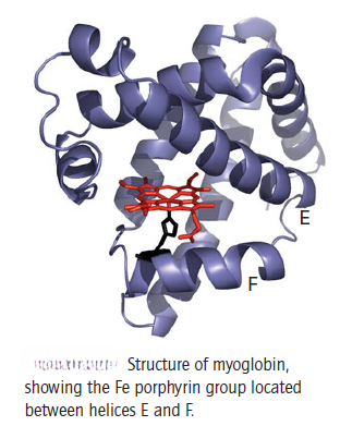
Myoglobin is an Fe protein that coordinates O2 reversibly and controls its concentration in tissue. The molecule contains several regions of alpha helix, implying mobility, with the single Fe porphyrin group located in a cleft between helices E and F. Two propionate substituents on the porphyrin interact with solvent H2O molecules on the surface of the protein. The fifth ligand to the Fe is provided by a histidine-N from helix F, and the sixth position is the site at which O2 is coordinated. In common terminology, the side of the haem plane at which exchangeable ligands are bound is known as the distal region, while that below the haem plane is known as the proximal region. The histidine on helix F is one of two that are present in all species. Such ‘highly conserved’ amino acids are a strong indication that evolution has determined that they are essential for function. The other conserved histidine is located on helix E.
Deoxymyoglobin (Mb) is bluish red and contains Fe(II); this is the oxidation state that binds O2 to give the familiar bright red oxymyoglobin (oxyMb). In some instances deoxymyoglobin becomes oxidized to Fe(III), which is called metmyoglobin (metMb) and is unable to bind O2. This oxidation may occur by a ligand substitution-induced redox reaction in which Cl- ions displace bound O2 as superoxide:
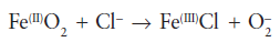
In healthy tissue, an enzyme (methaemoglobin reductase) is available to reduce the met form back to the Fe(II) form.
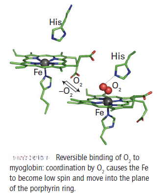
The Fe in deoxymyoglobin is five-coordinate, high-spin, and lies above the plane of the ring. When O2 binds it is coordinated end-on to the Fe atom, the electronic structure of which is tuned by the F helix histidine ligand. The unbound end of the O2 molecule is fastened by a hydrogen bond to the imidazole-NH of the histidine in helix E. The coordination of O2 (a strong-field π-acceptor ligand) causes the Fe(II) to switch from high spin (equivalent to t2g4 eg2) to low-spin (t2g6) and, with no d electrons in antibonding orbitals, to shrink slightly and move into the plane of the ring. The bonding is often expressed in terms of Fe(II) coordination by singlet O2, in which the doubly occupied antibonding 2πg orbital of O2 acts as a sigma donor and the empty 2πg orbital of O2 accepts an electron pair from the Fe. An alternative description is often considered, in which the bonding is expressed in terms of low-spin Fe(III) coordinated by superoxide, O2-. With this model, the formation of metmyoglobin by reaction with anions is a simple ligand displacement.
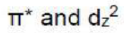
During the binding of O2 to myoglobin in the XY plane, sigma donation occurs from the dz2 orbital to the antibonding π* orbital of oxygen.