
Cell Cycle and Cell Division Neet Notes: Did you know that all organisms, regardless of size, begin as a single cell? You may marvel how a single cell can grow into such enormous animals. Cells, like all living entities, grow and reproduce. Cells multiply by dividing into two. Each parental cell produces two daughter cells. Daughter cells can multiply and divide, resulting in a new cell population from a single parental cell and its progeny. In other words, repeated cycles of growth and division enable a single cell to become a structure with millions of cells.
Cell Cycle and Cell Division Neet Notes
Read the complete explanation of the most important concepts of the NEET Class 11 Cell Cycle and Cell Division chapter –
What is Cell Cycle?
Cell division is an essential function in all living organisms. Cell division involves both DNA replication and expansion. Cell division, DNA replication, and cell development must be synchronized to produce healthy progeny cells with intact genomes. The cell cycle refers to the process of duplicating a cell’s genome, synthesizing its elements, and dividing into two daughter cells.
NOTE –
- The growth of cells (cytoplasmic increase) is continuous, although DNA synthesis happens only during one stage of the cell cycle.
- Cell division is a complex process of distributing replicated chromosomes (DNA) to daughter nuclei. These events are under genetic control.
Cell Cycle And Cell Division Handwritten Notes: Phases of the Cell Cycle
Human cells grown in vitro demonstrate a typical eukaryotic cell cycle.
Phases of the Cell Cycle
These cells divide about once every 24 hours. However, the period of the cell cycle varies by organism and cell type. Yeast, for example, can go through the cell cycle in approximately 90 minutes.
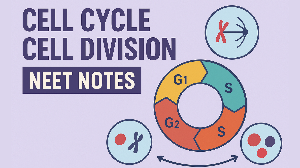
The cell cycle is broken into two main phases:
1. Interphase and
2. M Phase (Mitosis)
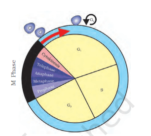
The M Phase is when cells divide or undergo mitosis, while the interphase occurs between two M phases. The average human cell cycle lasts 24 hours, with cell division lasting only approximately an hour. Interphase lasts over 95% of the cell cycle.
The M Phase begins with nuclear division, which separates daughter chromosomes (karyokinesis), and typically ends with cytoplasm division. The interphase, despite the name, the resting phase, is when cells prepare for division by growing and replicating DNA in an orderly manner.
The interphase is separated into three further phases:
- G1 Phase (Gap 1)
- S Phase (Synthesis)
- G2 Phase (Gap 2)
G1 Phase (GAP 1)
G1 phase occurs between mitosis and the start of DNA replication. During G1 phase, cells are metabolically active and expand continually, but do not duplicate their DNA. The S phase refers to DNA synthesis or replication. During this stage, the amount of DNA in each cell doubles. DNA starts at 2C and rises to 4C. Despite having diploid or 2n chromosomes at G1, the number of chromosomes remains constant after S phase, i.e. 2n.
What is the M Phase in the Cell Cycle?
During this stage of the cell cycle, the cell’s components undergo significant reorganization. Equational division occurs when the parent and progeny cells share the same number of chromosomes. Mitosis is separated into four stages of nuclear division (karyokinesis), although it’s important to note that cell division is a gradual process with no clear distinctions between stages. Karyokinesis has four stages:
The phases are:
- prophase,
- metaphase,
- anaphase, and
- telophase.
Cell Cycle And Cell Division Handwritten Notes For Neet
Learn the 4 most important step of the cell cycle here-
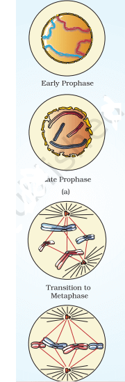
Prophase
Prophase, the first step of mitotic karyokinesis, occurs after the S and G2 interphases.
- In the S and G2 phases, new DNA molecules are interwoven rather than separate.
- During prophase, chromosomal material begins to condense. Chromosomes unravel during chromatin condensation.
- The centrosome, which was duplicated during the S phase of interphase, now moves to opposite poles of the cell. The completion of prophase is indicated by the following events:
- Compact mitotic chromosomes are formed by the condensation of chromatin. Chromosomes consist of two chromatids linked at the centromere.
- The centrosome, which was duplicated during interphase, begins to migrate to opposite poles of the cell. Asters are microtubules that sprout from each centrosome. The mitotic apparatus consists of two asters and spindle fibers.
- When seen under a microscope, cells at the end of prophase lack Golgi complexes, endoplasmic reticulum, nucleolus, and nuclear envelope.
Metaphase
The second phase of mitosis begins with the full collapse of the nuclear envelope, resulting in the spread of chromosomes throughout the cytoplasm.
- At this point, chromosomes are fully condensed and visible under a microscope. This stage is ideal for studying the shape of chromosomes.
- Metaphase chromosomes consist of two sister chromatids joined together by the centromere. Kinetochores are little disc-shaped structures found on centromere surfaces. These structures connect spindle fibers to chromosomes in the cell’s center.
- During metaphase, all chromosomes align at the equator, with one chromatid connected to spindle fibers from one pole and the sister chromatid connected to spindle fibers from the opposite pole.
- The metaphase plate refers to the plane where chromosomes align during metaphase. The major elements of metaphase include:
Spindle fibers are attached to chromosomal kinetochores.
Chromosomes migrate to the spindle equator and align along the metaphase plate via spindle fibers, reaching both poles.
Anaphases
During anaphase, each chromosome on the metaphase plate splits and produces two daughters.
- The chromatids, or daughter chromosomes of the future daughter nuclei, migrate towards opposite poles.
- As chromosomes travel away from the equatorial plate, the centromere stays at the front edge, while the arms trail behind .
- Thus, the anaphase stage is defined by major events that occur:
Centromeres divide, separating chromatids, which then migrate to opposite poles.
Telophase
During the final stage of karyokinesis, known as telophase, chromosomes at their respective poles decondense and lose their identity.
- Individual chromosomes are no longer visible, and chromatin material accumulates at both poles.
- This stage highlights the following major events:
Chromosomes cluster at opposite spindle poles, losing their identity as distinct pieces.
The nuclear envelope forms around chromosome clusters at each pole, generating two daughter nuclei.
Nucleolus, Golgi complex, and ER reform.

Cytokinesis – Separation Of Cytoplasm
Mitosis involves not only the separation of duplicated chromosomes into daughter nuclei (karyokinesis), but also occurs through cytokinesis, which involves the separation of cytoplasm into two daughter cells.
- Animal cells achieve this by forming a furrow in their plasma membrane. The furrow deepens and eventually merges in the center, separating the cell cytoplasm into two. Plant cells have an inextensible cell wall and so use a distinct cytokinesis mechanism.
- Plant cells produce walls from the center outward, meeting existing lateral walls. The creation of a new cell wall starts with a simple precursor called the cell plate, which is the median lamella between two adjacent cells. During cytoplasmic division, organelles such as mitochondria and plastids are dispersed between two daughter cells. In some organisms, karyokinesis does not progress to cytokinesis, resulting in a multinucleate state and the production of a syncytium, such as liquid endosperm in a coconut.
Significance of Mitosis
Mitosis, also known as equational division, typically occurs in diploid cells. However, in some lower plants and social insects, haploid cells divide through mitosis. Understanding the role of division in an organism’s life is crucial. Mitosis produces diploid daughter cells with identical genetic complements.
Mitosis is the process that drives multicellular organism growth. Cell development disturbs the balance between the nucleus and the cytoplasm. In order to reestablish the nucleo-cytoplasmic ratio, the cell must divide.
Cell Cycle And Cell Division Notes Class 11 Pdf
Sexual reproduction produces children through the fusion of two gametes, each with a complete haploid set of chromosomes.
Cell Cycle Cell Division Neet Notes Pdf Free Download – Click Here(Complete Notes)
What is Meiosis?
Gametes are generated from specialized diploid cells. Haploid cells are produced through specialized cell division, which reduces the number of chromosomes by half. This type of division is known as meiosis.
The major elements of meiosis are as follows:
- Meiosis has two separate phases of nuclear and cell division, meiosis I and meiosis II, but only one round of DNA replication.
- Meiosis I occurs after the parental chromosomes have replicated to form identical sister chromatids during the S phase.
- Meiosis pairs homologous chromosomes and allows for recombination between non-sister chromatids.
- Four haploid cells are generated at the end of meiosis II.
Meiotic occurrences can be divided into the following phases:
| Phases of Meiosis Cell Division | |
| Meiosis I Prophase I Metaphase I Anaphase I Telophase I |
Meiosis II Prophase II Metaphase II Anaphase II Telophase II |
Prophase I
The initial meiotic division’s prophase is often longer and more intricate than the mitotic prophase. It is further separated into five phases based on chromosomal behavior: Leptotene, Zygotene, Pachytene, Diplotene, and Diakinesis.
Metaphase I:
The bivalents line up on the equatorial plate. Microtubules from the spindle’s opposite poles bind to homologous chromosomes’ kinetochores.
Anaphase I.
The homologous chromosomes split, but sister chromatids stay connected at their centromeres.
Telophase I
The pair of cells occurs when the nuclear membrane and nucleolus resurface, followed by cytokinesis.
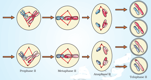
Meiosis II
This begins soon after cytokinesis, typically before the chromosomes fully lengthen. In contrast with meiosis I,
Meiosis II
Meiosis II mimics a typical mitosis. Figure 10.4 shows that the nuclear membrane dissolves at the conclusion of prophase II. The chromosomes again become compact.
Metaphase II
During metaphase II, chromosomes align at the equator and microtubules from opposite poles of the spindle bind to the kinetochores of sister chromatids (see Figure 10.4).
Anaphase II
Anaphase II begins with the splitting of each chromosome’s centromere, allowing sister chromatids to travel to opposing poles of the cell (Figure 10.4). This is achieved by shortening microtubules connected to kinetochores.
Telophase II
This stage marks the end of meiosis, when the two groups of chromosomes are covered by a nuclear envelope. Cytokinesis then occurs, culminating in the production of four haploid daughter cells.
Significance of MEIOSIS
Meiosis conserves a species’ chromosomal number across generations in sexually reproducing organisms, even though the process itself reduces chromosome quantity by half. This also leads to increased genetic heterogeneity among organisms throughout generations. Evolution relies heavily on variation.


 Karnataka 2nd PUC Result 2026 Date, Down...
 RSKMP Result 8th Class 2026 Declared @rs...
 Karnataka SSLC 10th Answer Key 2026 Rele...