
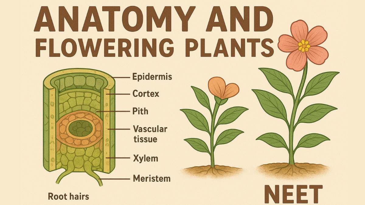
Anatomy And Flowering Plants NEET: The outward appearance of larger living organisms, including plants and animals, reveals structural similarities and variances. Similarly, when we study the internal structure reveal both similarities and variances. This Anatomy and Flowering Plants chapter of class 11 covers the internal structure and function of higher plants.
Anatomy is the study of plants’ internal structures. Plants are made up of cells, which are organized into tissues, and these tissues then form organs. Plant organs have distinct internal structures. Angiosperms exhibit anatomical differences between monocots and dicots. Internal structures also adapt to different conditions. In these Class 11 Anatomy of Flowering Plant Biology notes, we have provided simple explanations and summaries to break down difficult concepts.
Anatomy And Flowering Plants Notes Pdf
Class 11 NEET Biology Syllabus, Chapter 6 Anatomy and Flowering Plants Notes help students understand the interior anatomy of flowering plants. Important subjects covered in class 11 notes include plant tissues, roots, stems, and secondary growth. Examples and highlights of the complete Anatomy and Flowering Plants are given to get a clearer idea of the chapter.
Anatomy Of Flowering Plants Notes Class 11
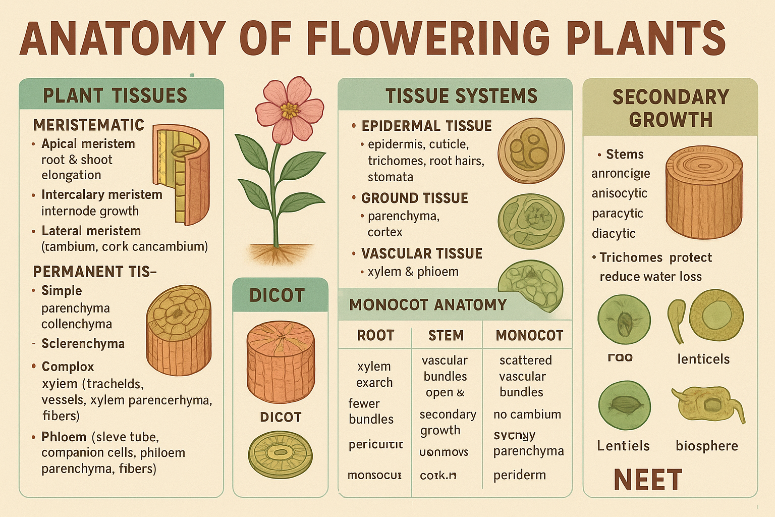
First, learn about all 3 main types of tissues of flowering plants –
Tissue System
We were discussing the different sorts of tissues based on the cells that were present. Let us now analyze how tissues differ based on their location in the plant body. Their structure and function vary according to locale. Tissue systems are classified into three groups depending on their structure and location. There are three tissue systems: epidermal, ground, and vascular.
1.Epidermal Tissue System
The epidermal tissue system covers the entire plant body and includes cells, stomata, and appendages such as trichomes and hairs. The epidermis is the outermost layer of a plant’s body. It consists of elongated, compactly packed cells that create a continuous layer. The epidermis is normally single-layered. Epidermal cells are parenchymatous, with a little quantity of cytoplasm on the cell wall and a big vacuole. The cuticle, a waxy layer on the epidermis, prevents water loss and protects the skin. The cuticle is lacking in roots. Stomata are structures found in the epidermis of leaves. Stomata control the process of transpiration and gas exchange. Each stoma consists of two bean-shaped cells known as guard cells, which enclose the stomatal outlet.
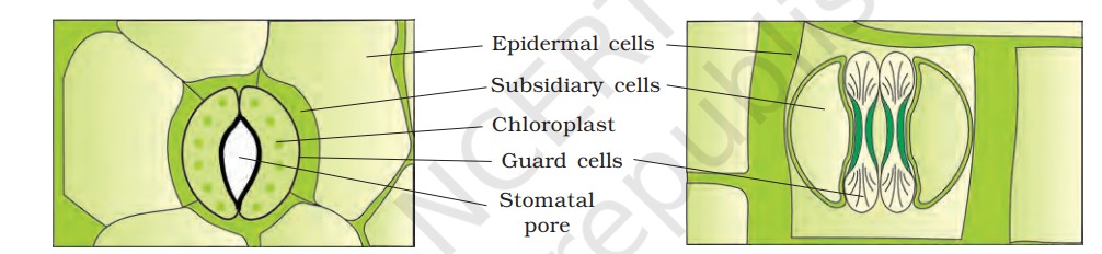
In grasses, guard cells are shaped like dumbbells. Guard cells have thin exterior walls (away from the stomatal hole) and robust inner walls. Guard cells have chloroplasts that regulate stomatal opening and shutting. Occasionally, a few epidermal cells near guard cells develop specialized shapes and sizes, referred to as subsidiary cells. Stomatal apparatus consists of the stomatal aperture, guard cells, and subsidiary cells.
The epidermal cells carry many hairs. Root hairs are unicellular extensions of epidermal cells that absorb water and minerals from the soil. Trichomes are epidermal hairs found on stems. The trichomes of the shoot system are typically multicellular.
2. Ground Tissue System
The ground tissue includes all tissues, excluding the epidermis and vascular bundles. It is made up of simple tissues, including parenchyma, collenchyma, and sclerenchyma. Parenchymatous cells are commonly found in the cortex, pericycle, pith, medullary rays, main stems, and roots. Mesophyll refers to the ground tissue of leaves, which consists of thin-walled chloroplast cells.
3. Vascular Tissue System
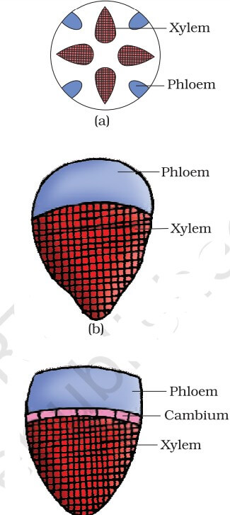
The vascular system is made up of complex tissues called phloem and xylem. Vascular bundles are made up of xylem and phloem. In dicotyledonous stems, cambium is found between the phloem and the xylem. Because of the presence of cambium, such vascular bundles can develop secondary xylem and phloem tissues, which is why they are known as open vascular bundles.
The vascular bundles in monocotyledons do not include any cambium. be they do not produce additional tissues, they are referred to be closed. Radial arrangement occurs when xylem and phloem within a vascular bundle are placed alternately along distinct radii, as seen in roots. In conjoint vascular bundles, xylem and phloem share the same radius. Such vascular bundles are seen in both stems and leaves. Phloem is typically found on the xylem’s outer side in conjoint vascular bundles

Anatomy Of Flowering Plants NCERT PDF
The anatomy of dicotyledonous and monocotyledonous plants.
Dicotyledonous and Monocotyledonous Plants
To better comprehend the tissue organization of roots, stems, and leaves, analyze transverse sections of their mature zones.
Dicotyledonous Root
The figure shows a transverse section of the sunflower root. The interior tissue structure is as follows:
Epiblema is the outermost layer. Epiblema cells often generate unicellular root hairs. The cortex has multiple layers of thin-walled parenchyma cells with intercellular gaps. The cortex’s innermost layer is called endodermis. It has a single layer of barrel-shaped cells with no intercellular gaps. Endodermal cells’ tangential and radial walls contain suberin, a water-impermeable waxy substance that forms Casparian strips. The pericycle is a collection of thick-walled parenchymatous cells located next to the endodermis.
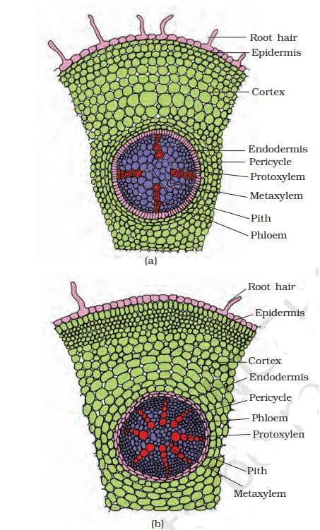
Monocotyledonous Root
In many ways, the monocot root resembles the dicot root. It consists of epidermis, cortex, endodermis, pericycle, vascular bundles, and pith. Monocot roots often have more than six (polyarch) xylem bundles, unlike dicot roots that have fewer. Pith is huge and well-developed. Monocotyledonous roots do not undergo secondary growth.
Dicotyledonous stem
A transverse section of a typical immature dicotyledonous stem reveals that the epidermis is the stem’s outermost protective layer. Trichomes and stomata may be present under a thin layer of cuticle. The cortex consists of cells grouped in layers between the epidermis and pericycle. It comprises of three sub-zones. The outer hypodermis is made up of collenchymatous cells underneath the epidermis, providing mechanical support to the young stem. Cortical layers beneath the hypodermis are made up of spherical, thin-walled parenchymatous cells with visible intercellular gaps. The innermost layer of the cortex is known as the endodermis.
Monocotyledonous stem
The monocot stem features a sclerenchymatous hypodermis, numerous vascular bundles surrounded by sclerenchymatous bundle sheaths, and a prominent parenchymatous ground tissue. Vascular bundles are connected and closed. Peripheral vascular bundles are often smaller than centrally situated ones. The absence of phloem parenchyma results in water-containing holes inside vascular bundles.
Dorsiventral (dicotyledonous) leaf
A vertical segment of a dorsiventral leaf through the lamina reveals three primary parts: epidermis, mesophyll, and the vascular system. The leaf’s upper (adaxial) and bottom (abaxial) epidermis have distinct cuticles. The abaxial epidermis often has more stomata than the adaxial epidermis. The latter may lack stomata. Mesophyll refers to the tissue between the upper and lower epidermis. Mesophyll, which contains chloroplasts for photosynthesis, is composed of parenchyma. It has two types of cells: palisade and spongy parenchyma.
Isobilateral (monocotyledonous) leaves
Isobilateral leaves share numerous similarities with dorsiventral leaves. It exhibits the following distinguishing differences. Isobilateral leaves have stomata on both surfaces of the epidermis and lack differentiation between palisade and spongy parenchyma.
In grasses, adaxial epidermal cells along veins transform into big, colorless cells. These are called bulliform cells. When the bulliform cells in leaves absorb water and become turgid, the leaf surface is exposed. Water stress causes flaccid leaves, which curl inwards to reduce water loss.
Anatomy Of Flowering Plants Short Notes For NEET PDF
The complete anatomy of plants is noted with detailed explanation, with key facts emphasized in Anatomy Of Flowering Plants Notes PDF to assist pupils in easily absorbing and recalling important knowledge. Download the pdf below for free –
Anatomy Of Flowering Plants Short Notes PDF Free Download – Click Here (Will be Updated Soon)


 Karnataka 2nd PUC Result 2026 Date, Down...
 RSKMP Result 8th Class 2026 Declared @rs...
 Karnataka SSLC 10th Answer Key 2026 Rele...