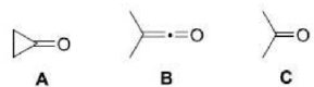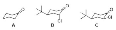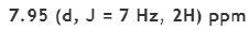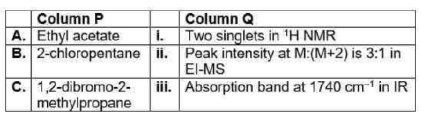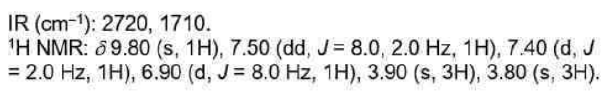
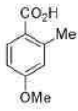
Correct option is C
Nuclear magnetic resonance spectroscopy, most commonly known as NMR spectroscopy or magnetic resonance spectroscopy (MRS), is a spectroscopic technique based on re-orientation of atomic nuclei with non-zero nuclear spins in an external magnetic field. This re-orientation occurs with absorption of electromagnetic radiation in the radio frequency region from roughly 4 to 900 MHz, which depends on the isotopic nature of the nucleus and increased proportionally to the strength of the external magnetic field. Notably, the resonance frequency of each NMR-active nucleus depends on its chemical environment. As a result, NMR spectra provide information about individual functional groups present in the sample, as well as about connections between nearby nuclei in the same molecule. As the NMR spectra are unique or highly characteristic to individual compounds and functional groups, NMR spectroscopy is one of the most important methods to identify molecular structures, particularly of organic compounds.
NMR-active nuclei, particularly those with a spin quantum number of 1/2, are of great significance in NMR spectroscopy. Examples include 1H, 13C, 15N, and 31P.
The energy difference ΔE between nuclear spin states is proportional to the magnetic field. ΔE is also sensitive to electronic environment of the nucleus, giving rise to what is known as the chemical shift, δ.
Shielded and Deshielded Protons
The valence-shell electron densities vary from one proton to another. In an applied magnetic field, i.e., when the molecule is placed in a uniform external magnetic field, the valence electrons around the protons are induced to circulate and this circulation, in turn, generates a secondary magnetic field, i.e., induced magnetic field. Circulation of electrons (specially πelectrons) about the nearby nuclei generates a field that can either oppose or reinforce the applied field at that proton. If the induced field opposes the applied field, the proton feels a lower field strength, and then the proton is said to be shielded. But if the induced field reinforces the applied field, the proton feels a higher field strength, and thus, such a proton is said to be deshielded.
Therefore, the different protons in a molecule do not have resonance at exactly the same frequency. This variability is due to the fact that the protons in a molecule are surrounded by electrons and exist in slightly different electronic (magnetic) environments from one another. The protons are shielded or deshielded by the electrons that surround them.
To generate an NMR spectrum, magnetic field strength (B0) is increased from left to right. The signal for the reference compound tetramethyl silane (TMS) appears at the extreme right of the spectrum with δ = 0 ppm. Due to the low electronegativity of silicon, the shielding of equivalent protons in TMS is greater than most of the organic compounds. Consequently, for most of the organic molecules, signals of the protons appear to the left of TMS signal in the NMR spectra. The more deshielded proton signals appear at the higher δ value, lower field (downfield) which is towards the left side of the plot. The more shielded proton signals appear at the lower δ value, higher field (upfield) which is towards the right side of the plot.
Spin-spin coupling
Structural assignment is often helped by the observation of the spin–spin coupling, which gives rise to multiplets in the spectrum due to interactions between nuclear spins. Spin–spin coupling arises when the orientation of the spin of a nearby nucleus affects the energy of another nucleus and causes small changes in the location of the latter’s resonance. A multiplet of 2I+1 lines is obtained when a spin-1/2 nucleus (or a set of symmetry-related spin-1/2 nuclei) is coupled to a nucleus of spin I. The coupling of the nuclear spins of different elements is called heteronuclear coupling. Homonuclear coupling between nuclei of the same element is detectable when the nuclei are in chemically inequivalent locations.
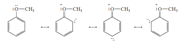
Electron-donating groups
A highly activating substituent such as methoxy clearly increases the electron density at the ortho and para positions of the ring (by resonance) and helps to give these protons greater shielding than those in the meta positions and thus a substantially different chemical shift.
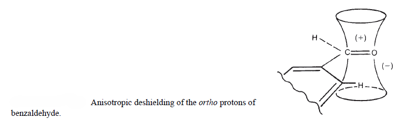
Anisotropy-Electron Withdrawing Groups
The ortho protons are much more deshielded than the meta and para protons due to the magnetic anisotropy of the π bonds in these groups. Anisotropy is observed when a substituent group bonds a carbonyl group directly to the benzene ring. Once again, the ring protons fall into two groups, with the ortho protons downfield from the meta/para protons.
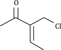
Proton (on the left) ortho to OMe will give a doublet at δ 6.90
Proton (on the left) ortho to CHO will give a double doublet at δ 7.50
Proton (on the right) ortho to CHO will give a doublet at δ 7.40