Correct option is C
Explanation -
¹H NMR with H/D exchange is a powerful tool to study backbone amide proton protection due to hydrogen bonding.
Different helical conformations (α, 3₁₀, π) have distinct hydrogen bonding patterns:
α-helix: i → i+4 hydrogen bonding
3₁₀ helix: i → i+3
π-helix: i → i+5
These variations affect how exposed or protected the backbone amide protons are from solvent. During H/D exchange, solvent-accessible amide protons exchange faster with deuterium.
Thus, NMR can detect which protons are protected, helping identify the type of helix present.
Option c - 1H NMR spectroscopy involving Hydrogen/Deuterium exchange
It directly measures the rate at which amide protons exchange with deuterium. Since α, 3₁₀, and π helices differ in their hydrogen bonding, the protection of amide protons will be distinct.
NMR gives residue-level information — you can tell which residues are in which type of helix. It is a quantitative and site-specific method.
Incorrect options-
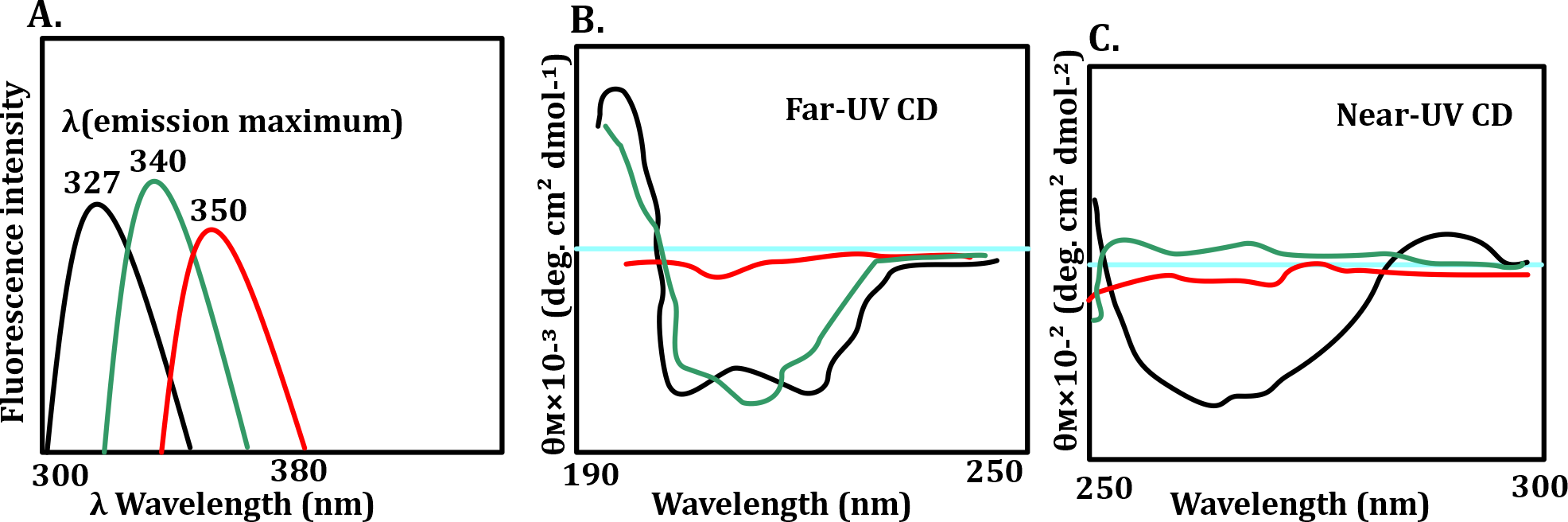
Option a - Near UV absorption spectrum between 250–300nm
Monitors aromatic side chains like Trp, Tyr.
Tells about tertiary structure, not backbone conformation.
Option b - Fluorescence emission spectra between 350–400nm
Again, mostly detects side chains like Trp.
Not useful for detecting helix type.
Option d - Near UV Circular Dichroism spectrum between 250–300nm
Far UV CD (190–250 nm) can distinguish α-helix vs β-sheet.
Near UV CD (250–300 nm) focuses on aromatic side chains, not backbone.
Even Far UV CD cannot distinguish α vs 3₁₀ vs π clearly.
So, the correct answer is option c - 1H NMR spectroscopy involving Hydrogen/Deuterium exchange