

Table of Contents
Human Eye
The human eye has a diameter of about 2.3 cm and is almost a spherical ball filled with fluid. It is made up of the following components: The sclera is the outer layer, a tough, protective white layer (white part of the eye)., while the cornea is the front, transparent portion of the sclera.
Human Eye Class 10
The Human Eye and Colourful World Class 10 Chapter 11 covers the following points:
- The accommodation of the eye is the capacity of the eye to focus on both close-up and distant objects by varying its focal length.
- The near point of the eye, or the least distance of distinct vision, is the shortest distance at which the eye can see objects clearly without exertion.
- It is roughly 25 centimetres for a young adult with normal vision.
- Myopia, hypermetropia, and presbyopia are three frequent refractive errors of the eye.
- By utilising a concave lens of the right power, myopia (short-sightedness, when the picture of distant objects is focussed before the retina) can be rectified.
- By utilising a convex lens of the appropriate power, hypermetropia (farsightedness, where the picture of adjacent objects is focused beyond the retina) can be rectified.
- With ageing, the eye’s ability to accommodate decreases.
- Dispersion is the division of white light into its individual colours.
- Light scattering is what gives the sky its blue hue and the Sun its reddish hue at sunrise and dusk.
DNA Full Form In Medical, Biology, English, Computer
Human Eye Definition
Human eyes are unique sense organs that can receive visual images and transmit them to the brain.
Human Eye Diagram For Class 10 Students
The Human Eye Diagram is very student for understanding the whole structure.
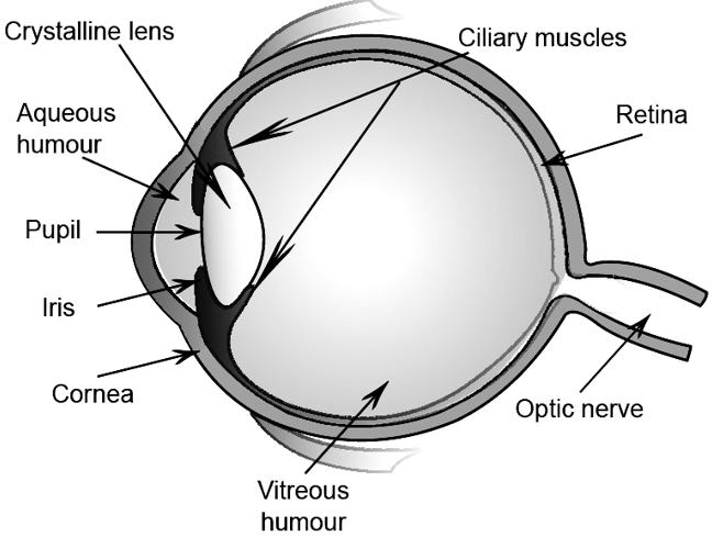
Human Eye Diagram
What Is Fungi?- Definition In Microbiology With Example
Eye Structure in Anatomy
Following is the anatomy of the Human Eye Structure:
- The orbit, which surrounds the eye and is built up of pieces of multiple skull bones to resemble a four-sided pyramid with its peak pointing back into the head, protects it from mechanical harm.
- The cornea, the eyeball’s front surface, must stay wet at all times.
- The eyelids help with this by periodically sweeping the lacrimal apparatus and other gland secretions over the surface during awake hours and covering the eyes during sleeping to keep moisture in and avoid evaporation.
- The conjunctiva borders the eyelids before bending back over the eyeball’s surface to form an outer covering for its anterior portion and ending at the cornea, the transparent section of the eye.
- The strong, relatively inflexible tarsal plates that border the palpebral aperture and the considerably thinner palpebral fascia, or sheet of connective tissue, make up the fibrous layer that gives the lid its mechanical stability; the two combined are referred to as the septum orbitale.
Structure of Eye for Class 10 Students
The human eye is a complex and remarkable organ responsible for vision. Its structure can be divided into several key parts:
- Cornea: The transparent outermost layer at the front of the eye. It helps in refracting light and protecting the eye.
- Iris: The colored part of the eye that surrounds the pupil. The iris controls the size of the pupil, regulating the amount of light entering the eye.
- Pupil: The black circular opening in the center of the iris. It allows light to enter the eye.
- Lens: Located just behind the iris, the lens further refracts light, focusing it on the retina.
- Retina: A thin layer of tissue at the back of the eye containing photoreceptor cells called rods and cones. These cells convert light into electrical signals, which are then sent to the brain through the optic nerve.
- Optic Nerve: A bundle of nerve fibers that connects the retina to the brain, transmitting visual information to the brain for processing.
- Sclera: The tough, white outer layer of the eye, covering most of its surface.
- Conjunctiva: A thin, clear membrane that covers the front surface of the eye and lines the inner surface of the eyelids.
- Aqueous Humor: The clear, watery fluid between the cornea and the lens, helping to maintain the eye’s shape and providing nutrients to the cornea and lens.
- Vitreous Humor: A gel-like substance filling the space between the lens and the retina, giving the eye its shape and helping to maintain its structure.
These components work together to allow light to enter the eye, focus on the retina, and convert visual information into electrical signals that the brain can interpret as images.
Human Eye lens
The lens’s primary optical purpose is to transmit light and focus it on the retina. The lens fine-tunes the focussing of light onto the retina, whereas the cornea contributes around 80% of total refraction. Although the human lens is white at birth, it gradually becomes more yellowish as people age33, which is likely because of the formation of 3-hydroxykynurenine and other tryptophan metabolites that filter UV radiation.
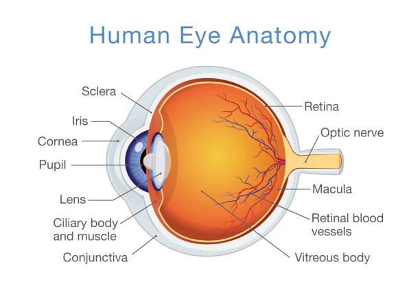
Human Eye Parts
The following are the parts of the human Eye:
- Cornea: the transparent part of the eye’s front that allows light to enter and concentrate (i.e., become sharp or clear). The cornea is reshaped during corrective laser surgery, which alters the focus.
- Pupil: The iris’s black, central opening is known as the pupil. The pupil enlarges or contracts depending on the amount of light present (smaller for bright light and larger for low light).
- Retina: the layer of nerves that lines the back of the eye. In response to light, the retina generates electrical impulses.
- Iris: the pigmented portion of the eye that aids in controlling light refraction. The iris shrinks the pupil to block out intense light when it is present. Additionally, the iris widens the pupil to let in more light when it is dark.
- Lens: Light rays are focused onto the retina via a lens. The transparent lens is removable if necessary.
- Optic Nerves: A bundle of more than a million nerve fibers known as the optic nerve carries visual information from the retina to the brain.
- Choroid: Between the retina, which is the inner light-sensitive layer, and the sclera is the choroid layer, which lines the back of the eye and contains blood vessels (the outer white eye wall).
- Macula: A region of the retina that is home to unique light-sensitive cells. These light-sensitive cells in the macula enable us to clearly discern little details in the middle of our visual field.
- Ciliary Body: Behind the iris, which focuses the lens, is a structure called the ciliary body, which contains muscle.
- Fovea: keen eyesight is provided by the macula’s centre.
- Sclera: the iris-surrounded, outer-white layer of the eye.
- Vascular Humor: The transparent, gelatinous material that lines the eye’s centre chamber.
Human Eye Structure and Function
The human eye is a complex organ that allows us to perceive our surroundings in the form of light and color. Here’s a brief overview of its structure and function:
Human Eyes Structure:
- Cornea: The transparent, front part of the eye that helps focus incoming light.
- Iris: The colored part of the eye, it controls the amount of light that enters the eye by changing the size of the pupil.
- Pupil: The dark, central opening in the iris that lets light into the eye.
- Lens: A transparent, biconvex structure situated behind the iris that further focuses light onto the retina.
- Retina: The light-sensitive layer of tissue at the back of the inner eye that contains photoreceptor cells (rods and cones).
- Rods and Cones: Photoreceptor cells in the retina responsible for vision in low light (rods) and color vision (cones).
- Optic Nerve: The nerve that carries visual information from the retina to the brain.
- Sclera: The white, outer layer of the eye that provides structural support.
- Choroid: A layer of blood vessels between the retina and the sclera, supplying the retina with nutrients.
- Vitreous Humor: The clear, gel-like substance that fills the space between the lens and the retina.
Human Eyes Function:
- Light Entry: Light first passes through the cornea, and then through the aqueous humor, pupil, and lens before reaching the retina.
- Focusing: The cornea and lens work together to focus light precisely onto the retina.
- Light Detection: The retina’s photoreceptors detect light and convert it into electrochemical signals.
- Signal Transmission: These signals are sent to the brain via the optic nerve, where they are interpreted as visual images.
- Adaptation: The eye can adapt to various light conditions by changing the size of the pupil and by adjusting the sensitivity of the rods and cones.
Also Read:
- All About Father Of Genetics- Gregor Mendel
- States And Union Territories Capitals Of India 2023
- 7 Rainbow Colours Name In Order, Drawing, Vibgyor Meaning
- What Is The Value Of Root 3?
- Which Is The Ugliest Language In India?
- How Much Is 1 Million And 1 Billion Dollars In Indian Rupees?
- What Is The Square Root Of 4?
- Smallest Country In The World By Area & Population, Map
- Compound Interest Formula, Definition, Calculator, Questions And Examples
- Unsung Heroes Of Freedom Struggle 1857 To 1947
 Types of Natural Resources with Examples
 All 100 Birds Name in Hindi and English
 50 Water, Aquatic, Ocean, Sea Animals Na...